(Upload on November 25 2018) [ 日本語 | English ]
Mount Usu / Sarobetsu post-mined peatland
From left: Crater basin in 1986 and 2006. Cottongrass / Daylily
HOME > Lecture catalog / Research summary > Glossary > Biology > Cell
|
Prokaryote: an organisms whose cells lack a membrane-bound nucleus (karyon)
Ex. yeast (monocellular), chlamydomonus, plants, animals (multicellular) Eukaryote: an organism whose cells contain a nucleus and organelles enclosed by membranes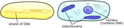 Typical cells of prokaryotes and eukaryotes Cell sizes
eukaryote (真核細胞) E. coli: 1 μm (1 × 3 or 1.5 × 3 μm) Streptococcus: 0.7 μm Chlamydia: 300 μm cowpox virus: 210 × 260 μm Major components of cellsprotoplasm (原形質)karyoplasm* (核質)
nuclear membrane (核膜)
cytoplasm (細胞質)
rough ER (粗面小胞体) |
Golgi apparatus (ゴルジ体) metaplasm (後形質)= the nonliving matter or inclusions, as starch or pigments, within a cellcell wall (細胞壁) vacuole (液胞) cell contents (細胞含有物) * karyoplasm, karyoplast, or nucleoplasm 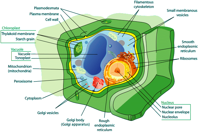 Plant cell structure |
☛ wood chemistry (木材化学)
|
Comparisons with animal cell → animal cells do not have cell walls Intercellular substance (細胞間物質) → Animal cell = no cellulose Plant cell = vegetal cellulose, hemicellular pectin, polysaccharide
glycocalyx (糖衣) = polysaccharide + glycocacidic polysaccharide Function
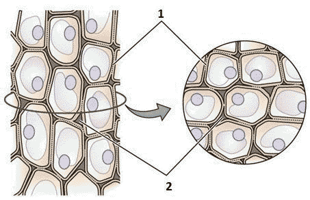
Cell divisionPlant cell: middle lamella (2 in Fig.) → primary wall (1)
Middle lamella: present between cells and acts as a cement preventing cells from migrating |
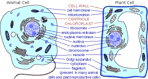 Fig. Animal and plant cells Plant cells are generally smaller than animal cells, due to vacuoles. Components
Cell fusion (細胞融合)cell → middle lamella (pectic subset, hemicellulose): working for cell adhesion→ primary wall → administrating cellulase and pectinase after pulling apart cells → protoplast, spheloplast → cell fusion 1978 Melchers (Germany)
Cell fusion of tomato (Lycopersicon esculentum Mill.) and potato (Solanum tuberosum L.) |
|
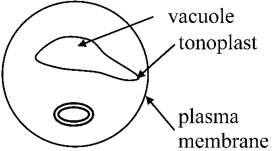
1776 Spallanzani: contractile vacuoles firstly observed in protozoa 1841 Dujardin: named vacuoles 1842 Schleiden: applied vacuoles for plant cells 1885 de Vries: named the vacuoule membrane as tonoplast A membrane-bound organelle that is present in all plant and fungal cells and some protist, animal and bacterial cells. |
|
|
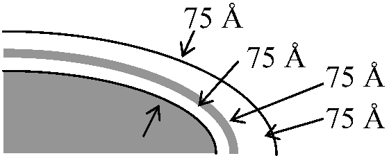
Unit membrane (単位膜) Plasma membrane makes a separation between vacuole and cytoplasm The structure of plasma membrane is similar with that of tonoplast 1895 Overtonthe membreans are perviousness to water and oil → the mmembrean consists of the mosaic of lipids and proteins 1917 Langmuir: stearate → monolayer1925 Gorter & Grende the fats developed on the membrane surface of red blood cells consist of two molecules or sheets 1935 Danielli & Davson:Sandwich model of plasma membrane 1950-60 Robertson
repeating unit theory(反覆単位説) unit membrane (基本膜構造) 1960 Neville: sundy punch of structure and function of membrane
isolated the liver plasma membrane of rats by homogenization
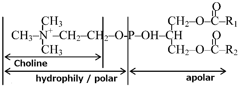
fatty substrates + phospholipids + α-lecithin + β-lecithin = 40%
sialic acid(シアル酸) and N-acethylneuraminic acid (NANA) are focused, because these substances are considered to be related to electrical characteristics of membrane surfaces 1964 Benzon
phase transition between protein and lipid → transition between crystal and liquid-crystal conditions negative staining of mitochondria by heavy metals → membrane consisting of protein-lipid complex 1972 Singer SJ & Nicolson GLfluid mosaic theory (流動モザイク説) 1974 Bretscher: utilized FMMP for probe (探針)
• impermeability through cell membranes
→ phosphatidyl ethanol amine is present in the inside of membrane
Phosphalidyl choline _________ 70-80% ___ foming outside the inside is also hydrolyzed when ghost cells are used
Outside __ SM ___ PC FMMP-35S 1980 Bretscher et al.
intact red dry ghost: proteins → gel electrophoresis
    Band MW         Weight
    No   (dalton)   %
    1    24         15.7 ___ pectin
    2    21.5       14.7 ___ pectin
    2.1  20         -    ___ ankyrin
    3    8.8-8.9    24.1 ___ protein band #3
    4.1  7.8        4.2
    4.2  7.2        5.0
    5    4.3        4.5 ___ actin
    6    3.5        5.5
    7    2.9        3.4
|
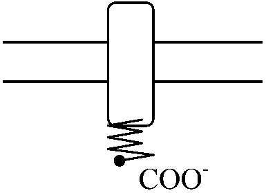
Ex. much glycophorin (sugar) - commassie blue Multiparticle model of cell membraneEnzymatic activityNa+-K+-ATPase: Ma++, Na+, K+ essential adenyl cyclase: 5'-ATP → cAMP (cell aggregation occrus after certaion cells of cellular slime molds discharge cAMP) 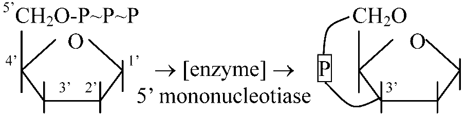
CH2-COR__________CH2OH sugar → glucose + fructose PeroxidaseMonophenol (-OH), polyphenol H2O2 + AH2 → 2H2O + A AA oxidase (amino-acid oxidase, アミノ酸酸化酵素)
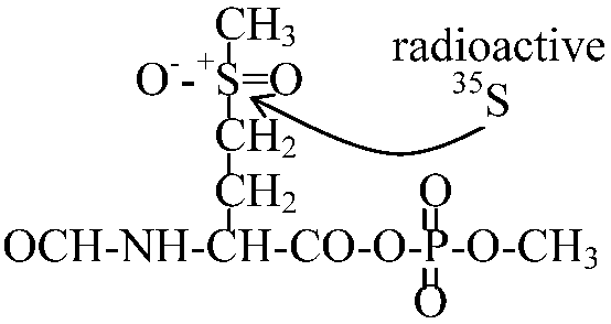
auxin(IAA) Selective permeability (選択的透過性)1952 Theorell
Giant axon(巨大軸索) of squid = giant nerve fiber
ATPase → the membrane is activated region =
1 molecule ATP 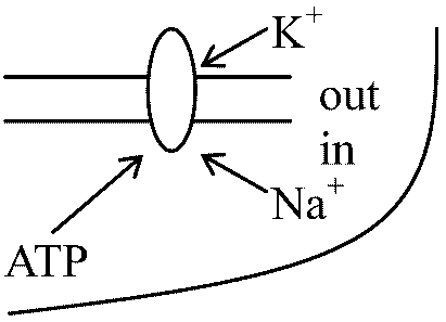
A-P-P-32P + Na+ (in) + E1(ATPase) → ATPase = 2.5-3.0 × 105 = subunits (large 1.0-1.3 × 10⁵) + (small 0.5 × 10⁵) 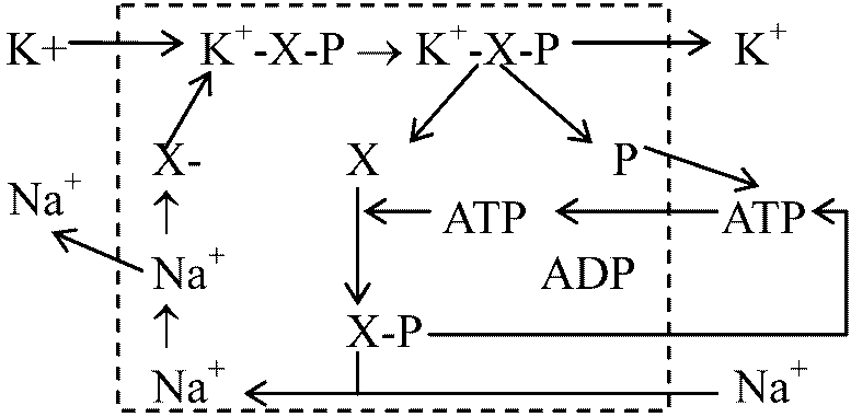
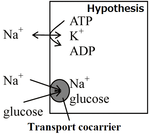
couple (共載) |
|
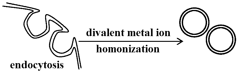
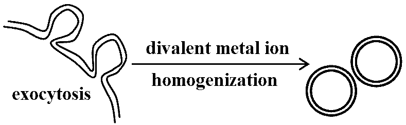
= protoplasmic membrane (formerly) 1970 Steck TL, Weinstein RS, Straus JH, Wallach DF
oragn or cell taking in nutrients → phagocytic vesicle |
|
|
1907 Veratti, Emilio: observed muscles
myofibrils enclosed by membrane structure 1945 Porter K: discovered endoplasmic reticulum =membrane structure within cells Rough-surfaced endoplasmic reticulum, rER (粗面小胞体)= granular endoplasmic reticulum (gER) or rough endoplasmic reticulum= containing ribosomes 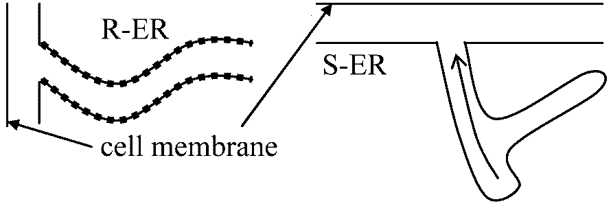 |
Smooth-surfaced endoplasmic reticulum, sER (滑面小胞体)= agranular endoplasmic reticulum (aER), smooth endoplasmic reticulum or smooth-surfaced endoplasmic= not containing ribosomes Microsomes (ミクロゾーム)heterogeneous vesicle-like artifacts (20-200 nm d) reformed from pieces of ER when eukaryotic cells are broken-up (in the laboratory)not present in viable and healthy cells |
Centrosome (中心体)Centriole (中心体)1. Spindle body → connected in fiber formation1956 Bernband: structure → globe → 5 μm = diameter of tublin sprial tube is formed by 13 tubulins are aggregated 2. Basal body
present in flagellate (鞭毛虫) and ciliate (繊毛虫) 9(3) + 2 → 9(3) + 3, 9(3) +1, 9(3) + 0 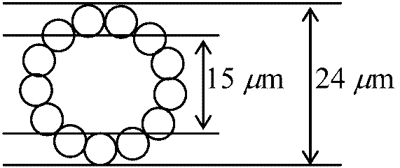 3. Sperm tail: 9+2 → common
4. Spindle fiber 1968 Miki & Sakai (三木・酒井)
spindle fiber consisting of microbubules (confirmed by electron microscope) |
1952 Mzaia & Dan
mitotic apparatus (紡錘体、中心体) → extracted from sea urchin and observed
Non-basic protein RNA Polysaccharide Spindle fiber consisting mostly of proteins (without DNA) 5. Microtubules
1963 Slantherbach: cnidoblasts of hydra 1952, 56 Loewy, Nakajima proteins in Physarum are actin (because of mobility) – rejected in the present 1953, 67 Kitching spirally lined-up microtubles → each microtuble consisting of 12 subunits? 1966 Nagai (永井), Rebhum: Chara 
50-100 filaments of which diameter is 50Å |
|
= Goldi apparatus, Golgi complex, dictyosome 1897 Golgi Camillo (Italian): identified the organella by a microscope
(called internal reticular apparatus) → |
FunctionPart of the cellular endomembrane system→ packages proteins into membrane-bound vesicles |
|
1897 Benda Carl (1857-1932): discovered mitochondria 1890 Altmann R: pointed out the similarities with bacteria 1900 Michaelis Leonor (1875-1949): janus green →strongly stained mt 1927 Wallin JE: mt originating from bacteria cell division: mt ≈ bacteria 1938 Bensley Robert R (1867-1956) & Bensley HS
Handbook of histological and cytological technique. University of Chicago Press
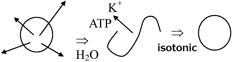
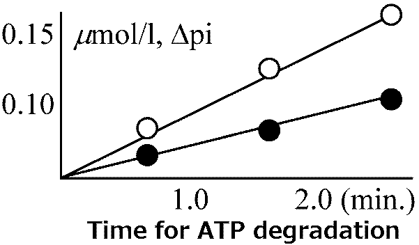
rat liver: 800 mitochondria/cell isolated mitochondria + Pi (リン酸) + ADP + NADH → ATP
increasing permeability by soaking into hypotonic solution
producing ATP → the measurements clarified (3 ATP)/NADH RC particles, supported fluid mosaic model 1945-50 Lehninger: isolated mitochondria
NAD: nicotic acid adenine dinucleotide (coenzyme I, DPN+): coenzyme cytochrome oxidase lacked in mt ≠ the gene in nuclei |
Structure 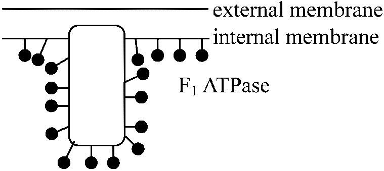
FunctionEnergy generatoroxygen (aerobic) respiration = ATP synthesis C6H12O6 + O2 → 12H2O + 6CO2 + 38ATP (688 kcal) TCA cycle: present in matrix ⇔ electron transfer system: present in cristae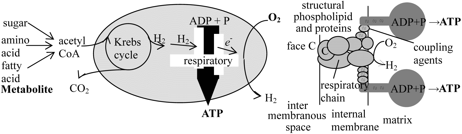 Fig. ATP synthesis Vesicles (小胞)1935 Szent-Györgyi: C4-dicarbonic acid cycle (now rejected)1937 Krebs: TCA cycle (クレブス回路, クエン酸回路) |
Plastid (色素体): chloroplast is a kind of plastidsChloroplast (葉緑体)a double-membrane-bounded organelle containing elaborated membranous sacs known as thylakoids; the membranes contain chlorophyll a and other components of the photpsynthetic light reactions → chlorophyll (葉緑素)
phaeoplast (褐色体) in brown algae and blue-green algae → fucoxanthin (褐藻素) Leucoplast (白色体): not containing chlorophyll Amyloplast (アミロプラスト): storing starch produced in chlorophyll Proplastid (プロプラスチド): un-developed pigments PhylogenyThe relationship between the phylogenetic development of photosynthetic apparatus and the system of classification in the plant kingdomEukaryotes (plastid)
Spermatophyta: Grana-and intergrana-thylakoid
Cyanophyta: Single or double thylakoid Structure1953 Frey-Wyssling & Steinmann
quantasome (etymology, quantum 光量) = ca 200 × 100 100 Å (80-100 Å)
regularly-arranged vesicles growing for four days in the darkness → lamella structure developed from prolamella body rapidly after exposure to light Stages: proplastid → differentiation → maturation Table. Dry weight (%) of chlorophyll content (Sinha 2004)Proteins _______ 35-55 (half in lamella) (Water soluble 20, Water insoluble 80)
Lipids _________ 20-30 (in lamella)  Granum (pl. -a): broadly defined as a stack(s) of thylakkoids within a chloroplast, such that the membranes of adjacent thylakoids are fused ☛ gene Expression of gene information in chloroplasts1883 Schimper & Meyer: Schimper and Meyer's theorythe concept of plastid continuity, stipulating that plastids do not arise de nove but from preexisting plastids (葉緑体有親説) |
1846 Näggeli:
all cells are coming from smaller cells = all chloroplasts are comoing from the chloroplasts
Nitella → mutants: the cells can not divide cells when the chloroplants are lost
proposed proplastid-origin theory → the structures of immature mitochondria are similar with the structures of proplastids considered that the chloroplasts are developed from the proplastids through deforming from immature mitochondria 1959 Stocking & Gifford
Spirogyra, 3H-thymidine radio autography - confirming that chloroplast has DNA Chloroplast – maternal inheritance (母性遺伝) Chloroplast RNARNA component: same with bacterial RNA1954 Jagendorf & Wildman, 1957 Chiba & Sugawara (千葉 & 菅原) confirmed that RNA is present in chloroplast 1962 Bandurskie & Maheshwari
14C-ATP: confirmed the synthesis of RNA polymerase
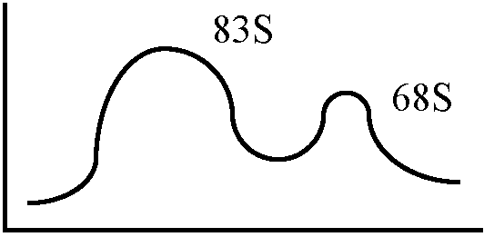
83S, 68S ⇒ sucrose density-gradient centrifugation - centrifuged high molecules
S: sedimentation coefficient (沈降定数), S-value, proposed by Sverdverg)
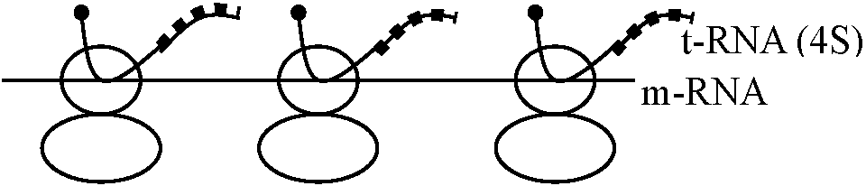
ribosome in leaf = 83S, 68S ↔ ribosome in root = only 83S enzyme: aninoacyl-t-RNA synthetase
a.a.-AMP-Et t-RNA → aminoacyl-t-RNA the presence of t-RNA and synthetase shown by adding 14C-valine and ATP 1964 Kirk: Vicia faba (ソラマメ) chloroplast
RNA containing 14C is synthesized when 14C-ATP, GTP, CTP and UTP are added - evidence on the synthesis RNA extracted from the chloroplast is 3.25S 1965 Gunning: EM = polymer → synthesize proteins
centrifugal separation of ribosomes from chloroplast just before and after exposing light
chlorophyll DNA: 40 μm
RNA polymerase is absent in chloroplasts |
Consisting of RNAc(60% RANs and 40% proteins in most cases)
Ribosome particles: small subunit + large subunit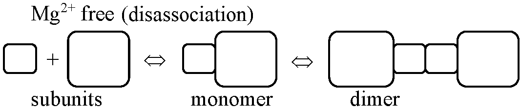 Small and large subunits (× 106, Chl. = chloroplast) E. coli Chl. Yeast Euglena Higher plants Large 1.1(23S) 1.1 1.24-1.30(25S) 1.35(24S) 1.30(25S) Small 0.56(16S) 0.56 0.6-0.7(17S) 0.85(20S) 0.70(17-18S) Mitochondrial ribosomeextracted from rat and Hela cells→ confirmed 50-60S ribosomes: 39-45S = large subunit (16S RNA), 25-35S = small subunit (12S RNA) |
cf. Yeast ribosome: 21S RNA + 15S RNA, Higher plants 26S + 18S 1971 Wu et al.
Hela's mitochondria → DNA, etc. → 16S RNA, 12S RNA Cytochrome b, cytochrome oxidase complex, oligomycin-sensitive ATPase
Chloramphenicol – mitochondria origin (inhibiting protein synthesis) RNA for producing enzymes related strongly to respiration (in mitochondria) are operated by nuculei, e.g.,malate dehydrogenase (MDH), isocitrate dehydrogenase (ICDH), β-hydroxybutyrate dehydrogenase, citrate dehydrogenase, and fumarate hydratase |
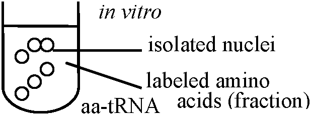
pl. nuclei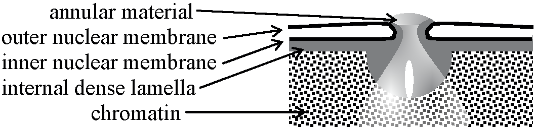 Fig. Nuclear membrane 1957 Allfrey, Mirskey, & Osawa: confirmed that proteins are synthesized in the nuculeus
Nucleolus (仁)Chromosomes of Trillium kamtscaticum
observed that nucleoli remained in the three parts chromosome duirng mid-stage after heat treatment |
Structurethe membrane of nucleolus can not be observed by EM1860 Manthver → nucleolanema 1956 Estalle ett al. → nucleolous
two different structures in nucleolus - two patterns recognized by dye staining Structural component1950 Casperson: condensed chromatin named as nucleolus-associated chromain 1952 Vincent
egg cells of starfish → Nuclear substances (核質)Nuclear substances or karyosome = chromonema (染色糸) + nucleolus (核小体)chromatintin, nuclear sap, nucleolus, nuclear skeleton, nuclear vacuole Chromatin (クロマチン) |
|
1848 Hofmeister W
discovered chromosomes in the dividing pollen mother cells of Tradescantia 1875 Strasburger: observed chromosome during cell division1879 Flemming described the splitting of dark stained rod-like structures during cell division which he called chromatin 1887 Beneden & Boveridiscovered that chromosome number in a species remains constant 1888 von Waldeyer-Hartz, Heinrich Wilhelm Gottfriedcoined the term chromosome (chromo- = colored, -some = body) 1902-03 Sutton & Boveri: proposed the chromosome theory of inheritance |
Fig. Chromosome. A, chromosome in metaphase of the cell cycle. The chromosome consists of chromatin, which is made of a molecule of DNA complexly coiled around a protein frame, forming a chromatid. Paired chromatids, consisting of identical molecules of DNA, are joined at the centromere. B, Normal male karyotype showing the 23 pairs of human chromosomes for a total of 46 arranged in 8 groups based on size and shape.
Salivary gland chromosomes (唾腺染色体)1881 Balbiani: discovered the giant chromosomepolytene chromosome (多糸染色体) Lumpbrush chromosome (ランプブラシ染色体)1882 Fremming W: discovered the chromosome |
|
1905 Mereschkowsky C: chloroplasts in higher plants
→ anucleate amebas cohabit 1918 Portier P: mitochondria are intracellular symbionts1927 Wallin IE: "Symbionticism and the origin of species" 1962, 63 Nass & Nass: colonial association theory
Structural and functional significance of bacteria and mitochondria 1969 Nass M: artificial symbiosis between mouse and chlorophyll |
 4. Both bacteria nebefit from the arrangement. 4. Both bacteria nebefit from the arrangement. 5. The internal bacteria are passed on from generation to generation. 5. The internal bacteria are passed on from generation to generation.Margulis, Lynn (1938-2011, Sagan = maiden name) 1967 Sagan L: "The origin of mitosing eukaryotic cells" proposed endosymbiosis (共生説) 1970 Margulis L: Origin of eukaryotic cells1970 Cohen SS |