(Upload on July 9 2025) [ 日本語 | English ]
Mount Usu / Sarobetsu post-mined peatland
From left: Crater basin in 1986 and 2006. Cottongrass / Daylily
HOME > Lecture catalog / Research summary > Glossary > Human anatomy
Terminologyfour-footed ↔ mankind (orthostatism, 直立位)
cranial (頭部) ↔ caudal (尾部) / superior (上側) ↔ inferior (下側) twins (双子), triplets (3つ子), quadruplets (4つ子), quintuplets (5つ子) quads (口), rigor mortis (死後硬直) Biopsy (生検)Def. a medical procedure in which a small sample of tissue, cells or fluid is removed from the body to be examined under a microscope by a pathologistto detect abnormalities such as cancer, inflammation, etc. Types: needle biopsy, punch biopsy, excisional biopsy, incisional biopsy, endoscopic biopsy, bone marrow biopsy, liquid biopsy |
☛ mankind, animal physiology |
Central nervous system, CNS (中枢神経系)Hollow brain (中腔脳) – vertebrate
effector = single innerevation 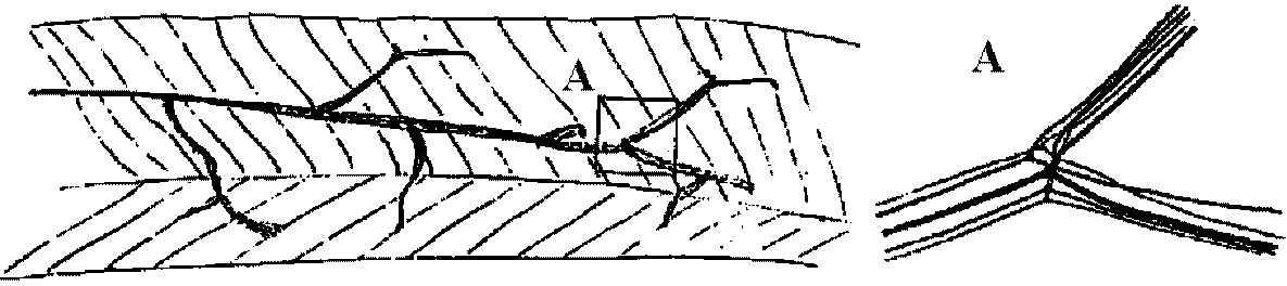 Fi.g 24.1. Sketch of the ramifications of the five axons (one inhibitory, four motor) innervating the flexor muscle of the rarpopodite of the lobster Panulirus. A shows a quintuplotomic branching enlarged. All muscle fibers are supplied by the branches of these five axons. (van Harreveld & Wiersma 1939) Uniterminal innervation________Dineuronal innervation Multiterminal innervation______Polynuronal innervation Fig. 7.2. Types of striated muscle innervation 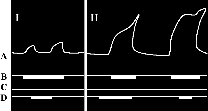
C = inhibitory motor neuron (inhibitor) 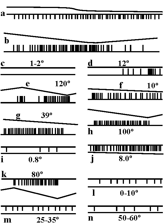 Fig. 18.8. Records of single units of the proprioceptor of the dactylopodire of Carcinus. The thin "line" indicates movement, up closing and down opening. (a) Pure position fiber with maintained discharge depending on the position of the joint. (b) Position fiber with sensitivity to the direction of movement. (c-h) Six different movement fibers with differing sensitivity. The numbers in (c-k) show the rate of movement in degrees per second. (i-k) One sensitive opening fiber, showing increase of the frequency at faster movement, which is shown in degrees per second. (i-n) The response of one sensitive closing fiber at three angles as shown, with a velocity of 1o/sec in all three records. (Wiersma & Boettiger 1959) 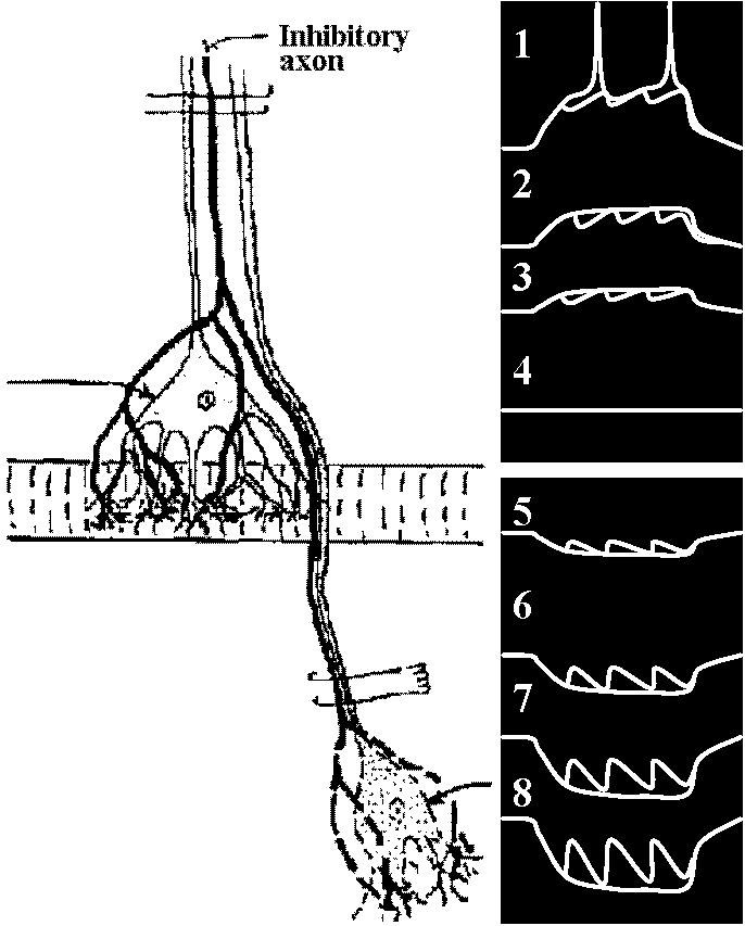 Fig. 20.17. Inhibitory postssynaptic potentials recorded with an intracellular microelectrode from a stretch receptor neuron of a crayfish (Aseacus). Left, a diagram of part of the preparation; two receptor neurons innervated by branches of one common inhibitory axon. The inhibitory axon is stimulated through a pair of electrodes applied near the "lower"' nerve cell. The action potentials travel along the inhibitory axon and reach the inhibitory synapses on the other nerve cell, where The inhibitory postsynaptic potentials (i.p.s.p.'s) are are being recorded at the site indicated by the arrow. (Since the original publication of this figure it has been found that inhibitory synapses are also located close to the recording site.) Right, a series of records (numbered 1 through 8) showing three i.p.s.p.'s each (each is preceded by a small vertical deflection, the stimulus artifact). Before the inhibitory axon was stimulated, the membrane potential of the sensory cell was artificially depolarized or hyperpolarized to different degrees, except in the ease of record 4. Note that the i.p.s.p.'s are hyperpolarizing during depolarization, and increasingly depolarizing during hyperpolarization of the membrane. The resting membrane potential happens to be equal to the reversal potential of the i.p.s.p.'s. (Left: Kniller 1955, Right: Hagiwara et al. 1960). 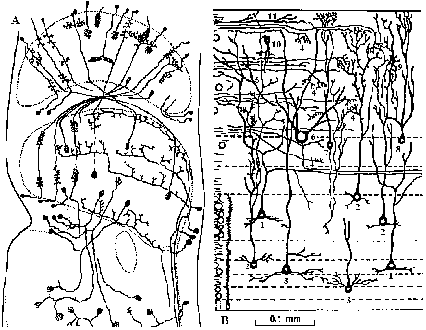 Fig. 2.53 Invertebrate vs vertebrate synaptic and nerve cell regions. A. Optic lobe of a crab. Golgi method. Only a selection of the cells and afferent fibers is shown. Note cell somas at periphery, without synapses; neuropile masses inside dotted lines include cell synapses but no cell somas (Hanstroem 1924). B. Optic lobe of a frog. Golgi method. Only a selection of the cells and afferent fibers is shown (the numbered cell types are described in Szekely et al. 1973); briefly they may be listed as: 1) large pyramidal neuron with type 1 dendrite. 2-3) large pear-shaped neurons with types 2-3 dendritic (adj. 樹木状の) arborization patterns. 4) optic nerve terminals. 5) ascending axon. 6) large ganglion cell. 7) small pear-shaped neuron with descending (adj. 下向きの) axon. 8) small pear-shaped neuron with beaded axon-like process. 9) stellate neuron (放射状ニューロン). 10) amacrine cell. 11) assumed endings of diencephalic afferent fiber. 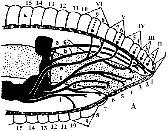 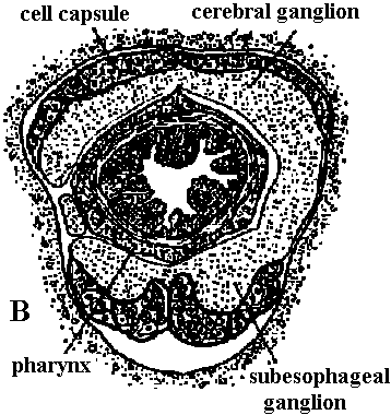 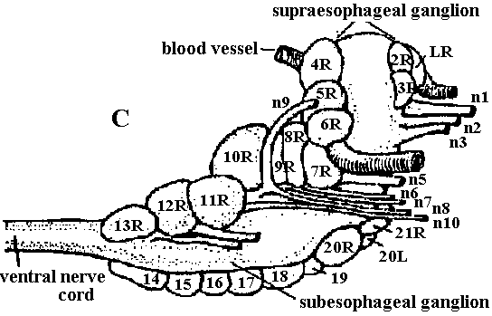 Fig. The anterior nervous system of leeches (吸血蛭, ヒル). A. Lateral view of the brain, subesophageal ganglion, and nerves of the head of Haemopis. Nerve roots are lettered o-f, annuli are numbered 1-15, segments 1-VI. B. Transverse section through the nerve collar, consisting of brain and subesophageal ganglion of Haemopis (Mann 1953). C. Lateral view of the brain of Theromyzon. n1-n12, cephalic nerves; cell compartments numbered L and R (Hagadorn 1958) 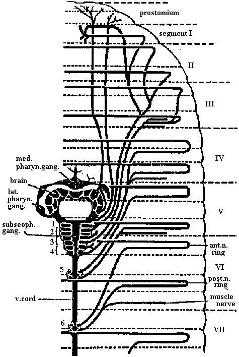 Fig. 14.6. Anterior end of the nervous system of Hirudo (蛭, ヒル), dorsal view. (Livanow 1904). ant.n.ring, anterior nerve ring; lat., med. pharyn. gang., lateral or median pharyngeal ganglion; post.n.ring, posterior nerve ring of the segment; subesoph. gang., subesophageal ganglion; v.cord, ventral cord; 1-6, presumed segments of the ganglia. CNS on humanCerebral cortex (大脳皮質)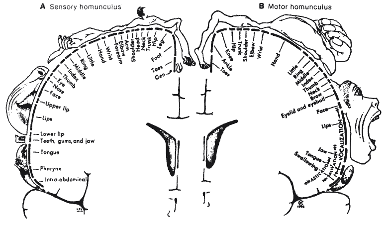 Fig. The motor and sensory homunculus in human brain shwoing the distribution of function role. (Penfield & Rasmussen 1950) Nerve system 12 pair cranial nerve (脳神経)
III-XII = cerebral nerves → I, II: not included in cerebral nerves (parts of brain) VertebrateA B 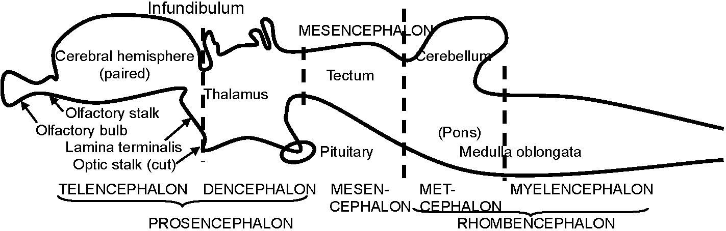 C Fig. 384. Diagrams to show the development of the principal brain divisions and structures. A, Only prosencephalon (primitive forebrain) distinct from remainder of neural tube. B, More mature stage in lateral view. C, The same in median section. |
Fig. 405. Diagram of the main centers and "wiring" arrangement of a reptile, in which the tectal region of the midbrain plays a dominant role; the corpus striatum (basal ganglia) is of some importance as a correlation center, but the neocortex (neopallium) is unimportant. The reticular formation of the brain stem (cross hatched) is important in carrying motor impulses to nuclei of the stem and cord. In this oversimplified diagram only a limited number of paths between somatic receptors and effectors are included; visceral centers and paths are omitted, as are cerebellar connections. Structure of basic brain stem (脳幹)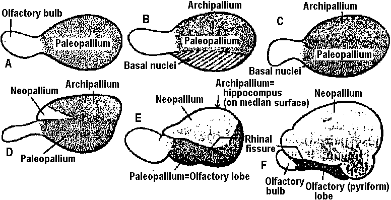 Fig. 400. Diagrams to show progressive differentiation of the cerebral hemisphere (cf. Fig. 401). Lateral views of left hemisphere and olfactory bulb. In A (primitive stage) the hemisphere is merely an olfactory lobe. B (Amphibian), Dorsal and ventral areas, archipallium (hippocampus) and basal nuclei (corpus striatum) are differentiated. C (primitive reptile), The basal nuclei have moved to the inner part of the hemisphere. D (advanced reptile), The neopalliurn appears as a small area (in many reptiles). E (primitive mammal), The archipallium is forced to the median surface, but the neopalliuin is still of modest dimensions, and the olfactory areas are still prominent below the rhinal fissure (as in primitive mammals). F (advanced mammal), The primitive olfactory area is restricted to the ventral aspect, and the neopallial areas are greatly enlarged (as in advanced mammals). The various cellular compounds of the hemispheres are distinguished by color. 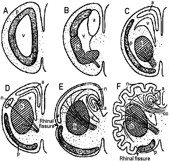 Fig. 401. Diagrammatic cross sections of left cerebral hemisphere to show stages in the evolution of the corpus striatum and cerebral cortex. A, Primitive stage, essentially an olfactory lobe; gray matter internal and little differentiated. B, St e seen in modern amphibians. Gray matter still deep to surface, but differentiated into paleopalli (= olfactory lobe), archipallium (= hippocampus), and basal nuclei (= corpus striatum), the last be ming an association center, with connections from and to the thalamus (indicated by lines representing cut fiber bundles). C, More progressive stage, in which basal nuclei have moved to interior, and pallial areas are moving toward surface. D, Advanced reptilian stage; beginnings of neopallium. E, Primitive mammalian stage; neopallium expanded, with strong connections with brain stem; archipallium rolled medially as hippocampus; paleopallial area still prominent. F, Progressive mammal; neopallium greatly expanded and convoluted (包旋形・回旋状); paleopallium confined to restricted ventral ar a as pyriform lobe. The corpus callosum developed as a great commissure (接合部, 交連部) connecting the two eopallial areas. a, Archipallium; b, basal nuclei; cc, corpus callosum; n, neopallium; p. paleopallium; V, ventricle. The different types of "gray matter"colorcd as in Fig. 400. 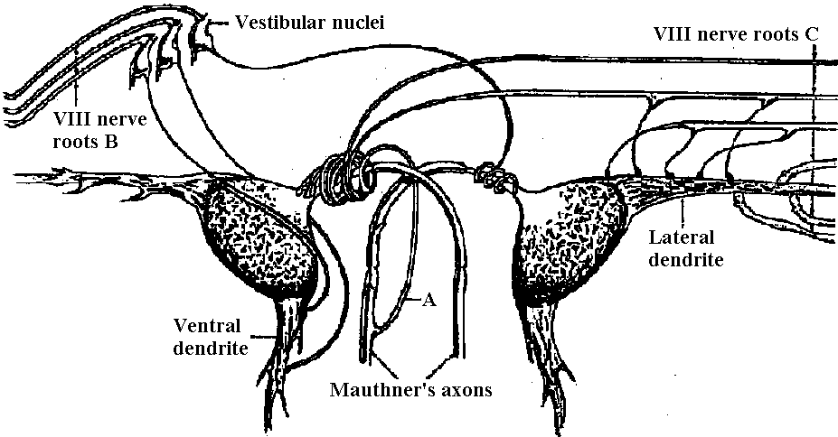 Fig. 7.13. A reciprocally inhibiting pair of neurons. The giant cells of Mauthner in the medulla of fish, schematically shown, with the inhibitory collateral (A) indicated only on the left, the indirect VIII nerve afferents (B) only on the left, and the direct VIII nerve afferents (C) coming only from the right, although all these components are really bilateral (Retzlaff & Fontaine 1960) 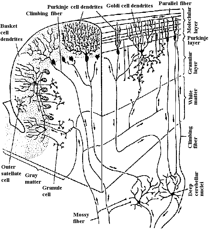 Fig. 3.15. Stereodiagram illustrating the principal neuronal types in the cerebellar cortex, the two afferent systems, and the interconnections between these elements. The mossy fiber afferents innervate granular cells and Golgi-cells in complex synaptic glomeruli. The granular cell axons then rise toward the surface of the cerebellum, where they branch to form parallel fibers that excite Purkinje cells, Golgi cells, basket cells, and stellate cells. The Purkinje cell axons constitute the only output from the cerebellar cortex. (Eccles et al. 1967) Medulla oblongata (延髄)Basic structure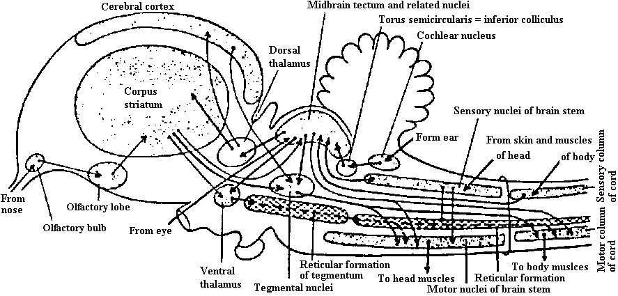 Fig. 406. A "wiring diagram" of a bird brain, comparable to that of Fig. 405. The midbrain tectum is still of importance, but the corpus striatum is the dominant center in many regards. 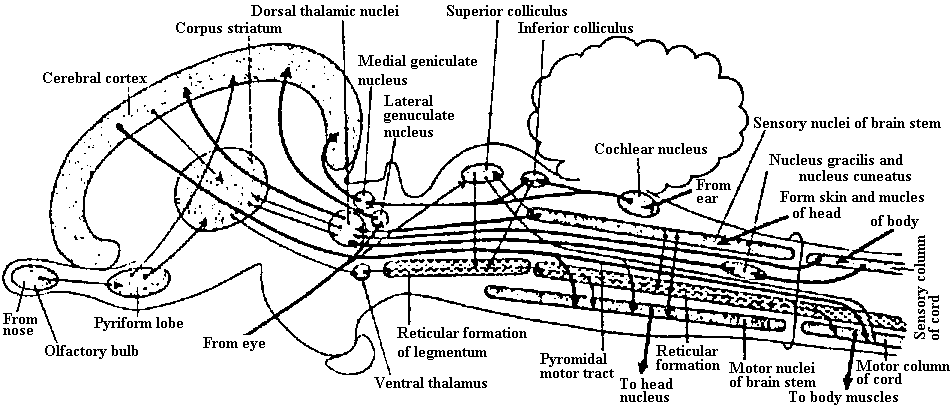 Fig. 407. A 'wiring diagram" of a mammalian brain comparable to Figs. 405 and 406. The midbrain tectum is reduced to a minor reflex center, and the corpus striatum is relatively unimportant; most sensory impulses are projected “upward” to the cerebral cortex, whence a direct motor path (pyramidal tract) extends to the motor centers and cord. Hindbrain (後脳)A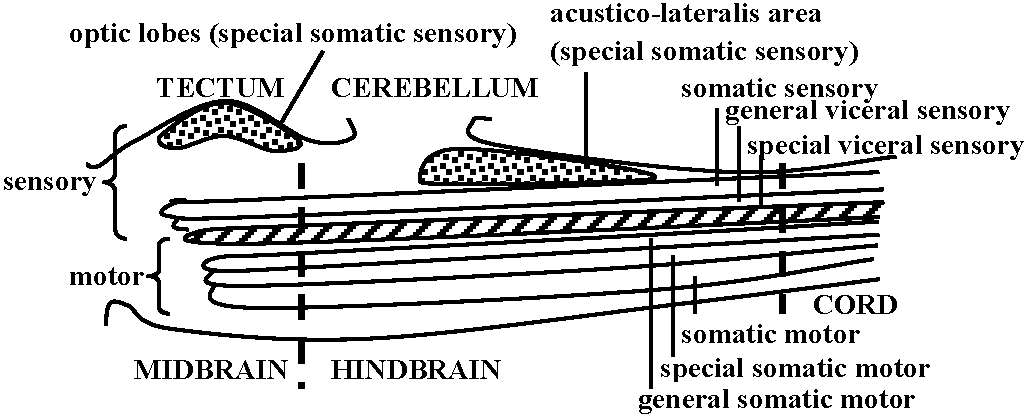 B 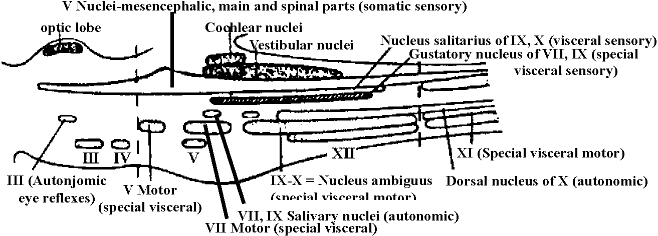 Fig. 398. Diagrams of midbrain and hindbrain regions in lateral view to show the arrangement of sensory and motor nuclei. Somatic sensory, blue;special somatic sensory, stippled; visceral sensory, green; special visceral sensory, hatched; visceral motor, yellow; special visceral motor, stippled; somatic motor, red. A, Hypothetic primitive stage, in which brain stem centers were continuous with one another and with the columns of the cord. Even at such a stage, however, it would be assumed that special somatic centers would have developed for eye and ear. The brain region includes a special visceral motor column for the branchial muscles. B, Comparable diagram of the mammalian situation. The somatic sensory column is still essentially continuous (almost entirely associated with nerveV), but the other columns are broken into discrete nuclei. The visceral sensory column includes both a general visceral nucleus (mainly for afferent fibers from the viscera via the vagus) and a special nucleus for the important sense of taste. Of efferent visceral nuclei, there are small anterior ones for automonic eye reflexes and the salivary glands, and a large nucleus for parasympathetic fibers to the viscera via the vagus. There are important branchial motor nuclei for V, VI, and IX, X (ambiguus). The somatic motor column includes small nuclei for eye muscles anteriorly and a hypoglossal nucleus posteriorly. Higher brain (高次脳)
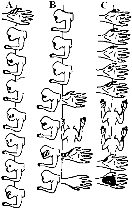
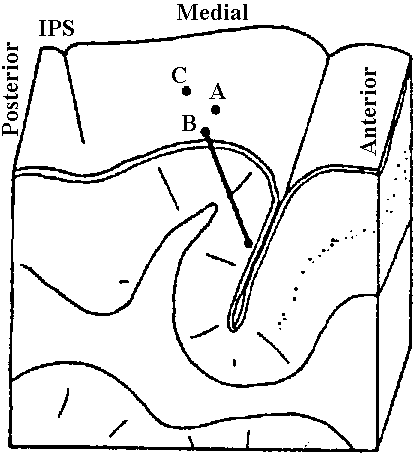
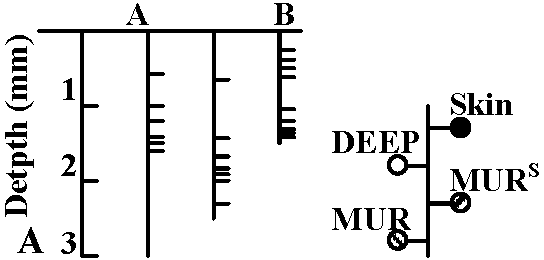
 Topographic organization of the somatosensory cortex of a primate, Macaca mulatta (IPS, intraparietal sulcus). A. Reconstruction of three microelectorde penetrations, at A, B, and C, through the cortex of the right hemisphere in a region calld S1 (first somatic area). The modality and receptive field (RF) of each neuron encountered is marked on the representations of body parts. The positions of the neurons along the penetration are indicated at the lower left: broken lines mean a multiunit record (MUR), solid lines a single unit (symbols indicate stimuli to the skin (S) or to deep cutaneous sense organs (o). All neurons depicted were responsive to hair movement or mechanical stimuli on the skin. Such detailed maps permit the following overall map. B. The postcentral gyrus is shown schematically unflolded, with the level of spinal origin of input shown to the left. Note the regular progression from face and head on the ventral-lateral surface to the tail at the top of the cortex, and the distortion of the map that results from emphasizing the analysis of input from the face, hands, and feet. da, dorsoaxial line; va, ventroaxial line; am, anterior midline; pm, posterior midline. The numerals (1, 2 , 3, 3a) along the cut lateral edge of the map indicate the cytoarchitectural fields that make up S1. (Whitsel et al. 1971) Lower brain (低次脳)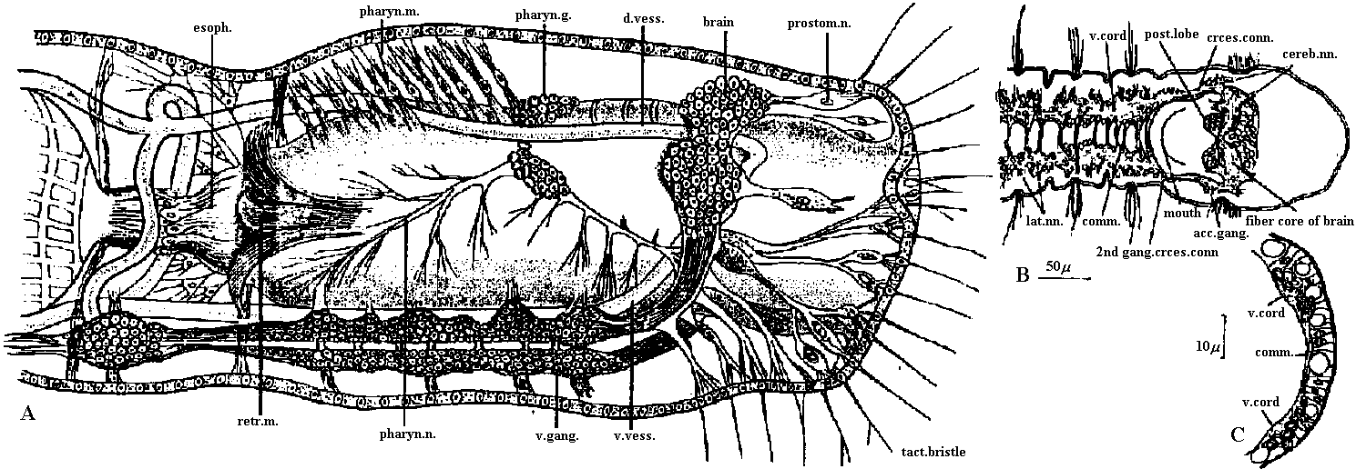 Fig. 14.5. The central nervous system of two lower oligochaetes. A. Chaetogaster, lateral view of anterior end. (Vejdovsk 1884). B. Aeolosoma, frontal section of anterior end. C. The same, transverse section of midventral epidermis, ventral to the right (Brace 1961). acc.gang., accessory ganglion; cereb.nn., cerebral nerves; comm., commissure; crces.conn., circumesophageal connective; d.vess., dorsal blood vessel; esoph., esophagus; 2nd gang.crces.conn., second ganglion on circumesophageal connective; lot.nfl., lateral nerves; pharyn.g.m.n., pharyngeal ganglin, muscle, and nerve; post.lobe, posterior lobe of brain; prostom.n., prostomial nerve; retr.m., retractor muscle; tact.bristle, tactile bristle; v.cord, ventral nerve cord ; v.gang., ganglion of ventral cord ; v. vess., ventral vessel. Human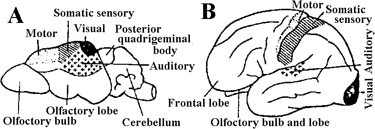 Fig. 404. Lateral view of A, the brain of a shrew (トガリネズミ); B, the cerebrum of man; to show cortical areas. Commissural fiber that connects the right and left hemispheres of the brain is larger in women than in men. Blood transfusion science (輸血学)1667 Denys, Jean-Baptiste (1643-1704): transfusion (sheep to human)eleven internal hemorrhaging patients - 6 of them survived 1818 Blundell, James (1791-1878): transfusion (human-to-human) |
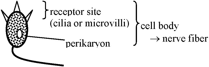
Anatomical classification1. Primary sensory cell (receptor)
Stimulus →
neurite (axon) = action potential → central nervous system Ex. vertebrate's olfactory organ, photoreceptor 2. Secondary sensory cell (receptor)
Vertebrates only Ex. ear lateral ine (acoustico-lateralis), taste bud
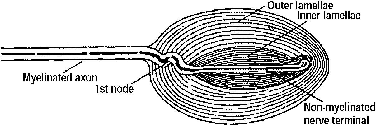
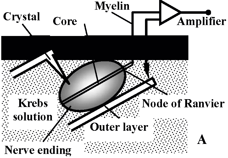
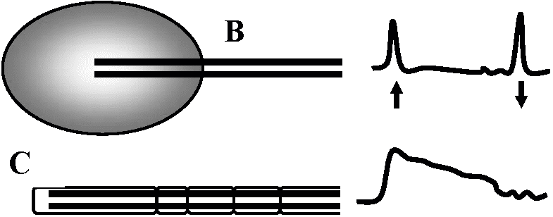
Pacinian corpuscle: given pressure – mammalian skin receptor
Meisner corpuscle: tactile skin – accessary cells
Krasne corpuscle: feeling coldness
Acoustic sense and taste sense (聴覚器・味覚器)Auditory apparatus (聴覚器)External ear (外耳)protection of inner ear from external laud sound attention of self-stimulus Middle ear, tympaum (中耳) Inner ear (内耳) 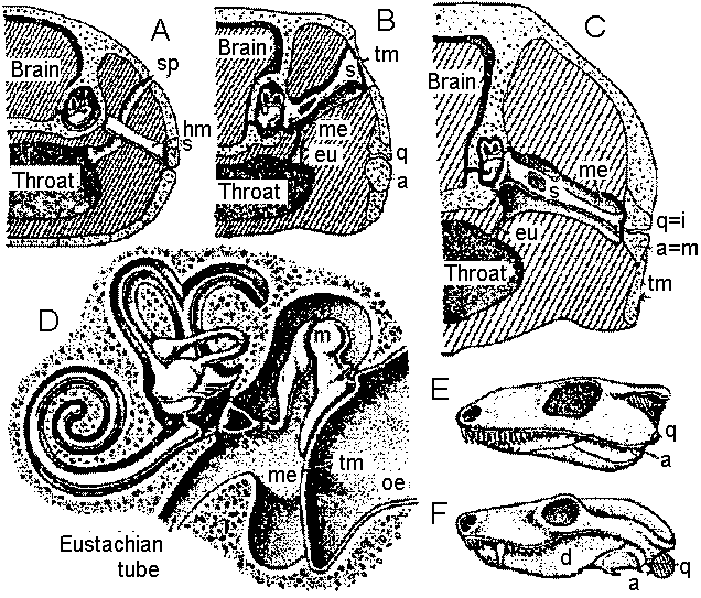 Fig. 359. Diagrams to show the evolution of the middle ear and auditory ossicles. Diagram. matic sections through the otic region of the head of A, a fish; B, a primitive amphibian; C. a primitive reptile; D, a mammal (showing the ear region only); E, side view of the skull of a primitive land vertebrate; F, of a mammal-like reptile to show the shift of the eardrum from tympanic notch (V字型の切り込み) of the skull to the region of the jaw articulation, a, Articulac; d, dentacy; eu, eustachian tuhe; boa, hyomandibular; i, incus; m, malleus; me, middle ear cavity; ne, outer ear cavity; q, quadrate; a, stapes; ap, spiracle; tin, tympanic membrane. (Romer, Man and the Vertebrates, UCP) 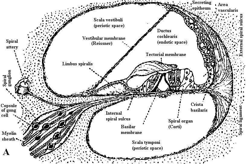 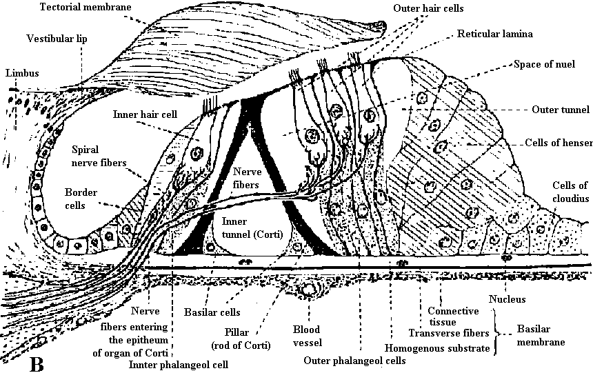 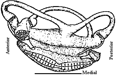
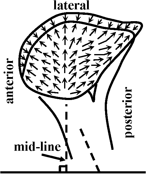 ____Fig. 14.___________________________Fig. 15 Fig. 14. Schematic drawing of the fish labyrinth (Lota vulgaris) demonstrating the morphological polarization in the various sensory epithelia of the labyrinth. The arrow indicates the direction of orentation of the sensory hairs as indicated by the location of the kinocilium. Fig. 15. Schematic drawing of orientation of sensory cells in the macula utriculi in the fish (Lota vulgaris). (Flock 1964) 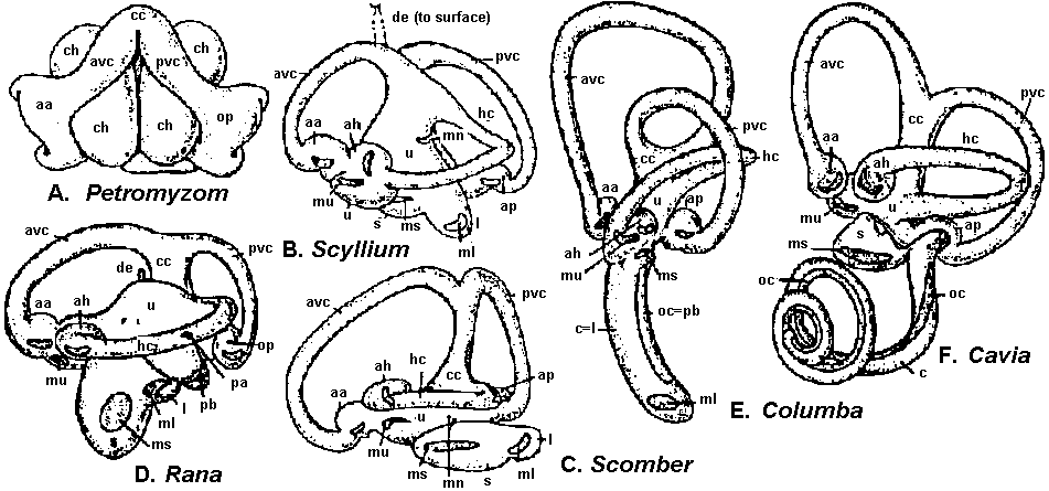 Fig. 355. Membranous labyrinth of A, lamprey; B, shark; C, teleost; D, frog; E, bird; F, mammal; all external views of the left ear. Sensory areas are shown (except in A) as if the membrane were transparent. aa, Ampulla of anterior canal; ah, ampulla of horizontal canal; ap, ampulla of posterior canal; aec, anterior vertical canal; c, cochlcar duct; cc, crus commune with which both vertical canals connect; ch, chambers in the lamprey ear lined with a ciliated epitheliuns; de, endo-lymphatic duct; hc, horizontal canal; I, lagena; in), macula of lagena; mu, macula neglecta; ma, macula of sacculus; mu, macula of utriculus; or, organ of Corti; pa, papilla amphibiorum; pb, papilla basilaris; pvc, posterior vertical canal; a, sacculus; u, utriculus. (After Retrius.) 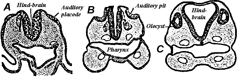  |
Fig. 356. Diagrams to show the development of the internal ear in mammals. A to C, Cross sections of the head of early emhryos; an ectodermal placode sinks inward on either side to form an otic vesicle. D to C, Successive stages in the development of the various parts of the memhranous labyrinth from the otic vesicle: c, cochlear duct; de, endolymphatic duct; sac, sacculus; ut, utriculus, (A to C from Arey; D to F after His and Bremer.)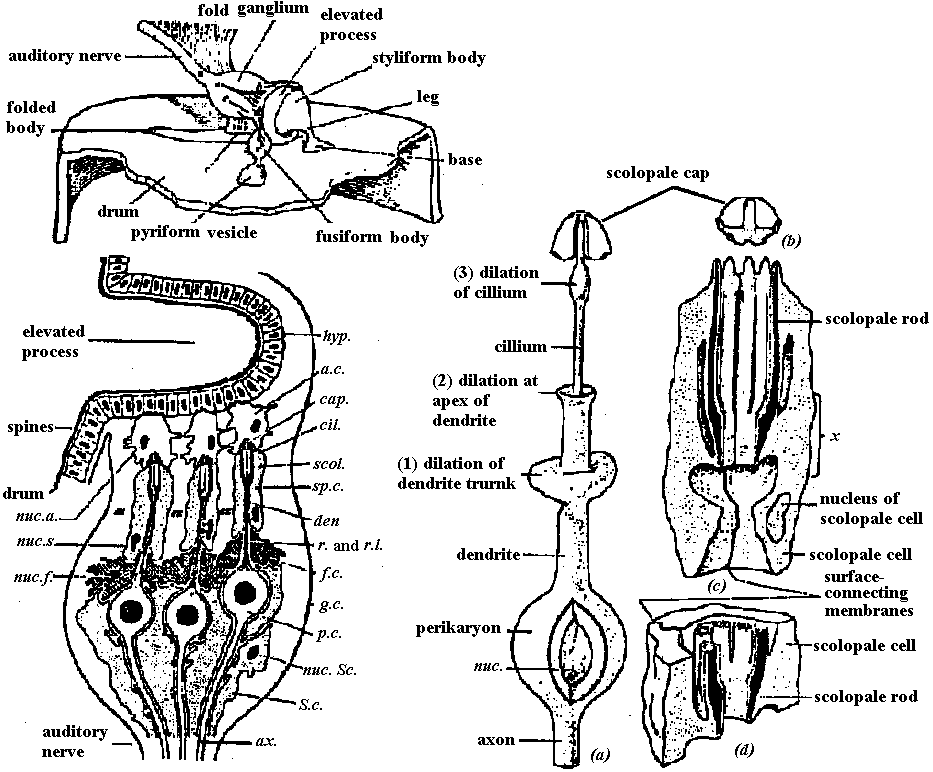 Fig. 25.1. Fine structure of the insect ear as revealed by electron microscopy of the ear of the locust (Locusta migratoria). The eardrum, or tympanic membrane, is located laterally on the first abdominal segment. The auditory ganglion is attached to its inner surface. It contains 60 to 80 sensory neurons. Their perikaryon (p.c.) and axon (ax.) are surrounded by Schwann cells (S.c.); one Schwann cell enfolds several sensory neurons. The proximal part of the dendrite is enclosed by a fibrous sheath cell (f.c.); the distal part of the dendrite (den) is surrounded by another type of satellite cell, the scoloplle cell (sp.c.). A cilium (cil.) extends from the dendrite into a scolopale cap (cap.). This in turn is in rontaet with an attachment cell (a.c.) that connects with the hypoclermal cells (hyp.) of the elevated process of the tympanic membrane. The root (r.) and rootlets (r.l.) of the cilium can be seen inside the dendrites. Note the extracellular flied space (es) between the scolopale cells. The nature of the mechanical deformation of the sensory neurons during vibration of the tympanic membrane is still unknown; but there is no doubt that these ears permit the female locusts to hear the “songs” of the courting males. g.c., capsule of ganglion; nur.a. nucleus of attachment cell; nuc.s., nucleus of scolopale cell; nuc.f., nucleus of fibrous sheath-cell; nuc.Sc., nucleus of Schwann cell; scol., scolopale. (Gray 1961) Gustatory organ (味覚器)= tongue (human)Visual sense (視覚)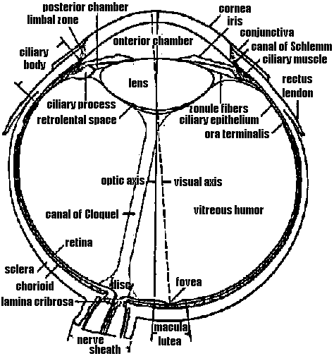 Fig. 1. Horizontal section of human eye. (Walls 1942) iris (虹彩) sclera (強膜) chorioidea (脈絡膜) macula lutea (黄点), centrocecal scotoma (盲点) retina (網膜) anterior chamber (前房) lens (レンズ) cornea (角膜) ciliary body (毛様体) optic axis (眼球軸) visual axis (視軸) fovea (網膜中心窩) pupil (瞳孔) 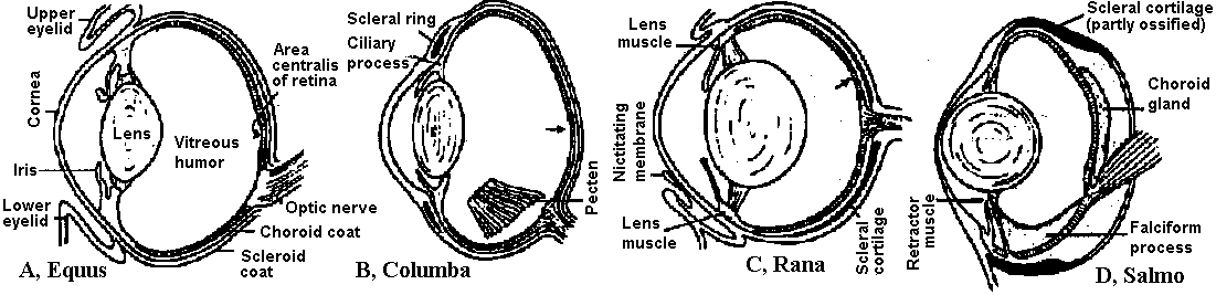 Fig. 345. Diagrammatic vertical sections through the eye of A, a horse; B, a dove; C, a common frog; D, a teleost (salmon). Connective tissue of sclera and cornea unshaded; scleral ring or cartilage black; choroid, ciliary body, and iris stippled; retina hatched. Arrows point to the fovea. In B is shown the pecten, lying to one side of the midline. In D the section is slightly to one side of the choroid fissure through which the falciform process enters the eyeball. 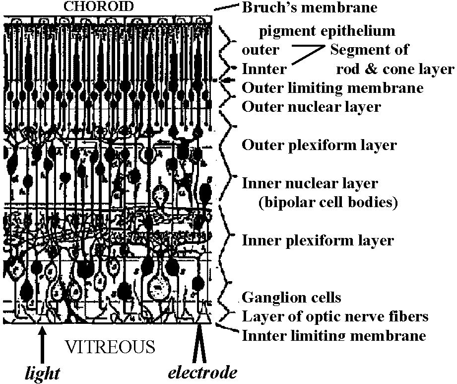 Fig. 12. Primate retina based on Golgi impregnation, showing principal cell types and their synaptic relations. Various cell types are: c, horizontal cells; d, e, f, diffuse or polysynaptic bipolar cells; h, individual cone (midget) bipolar cell; i, l, “amacrine cells”; m, n, o,p, r, s, ganglion cells, of which s is the individual or monosynaptic ganglion cell. (Polyak 1941) 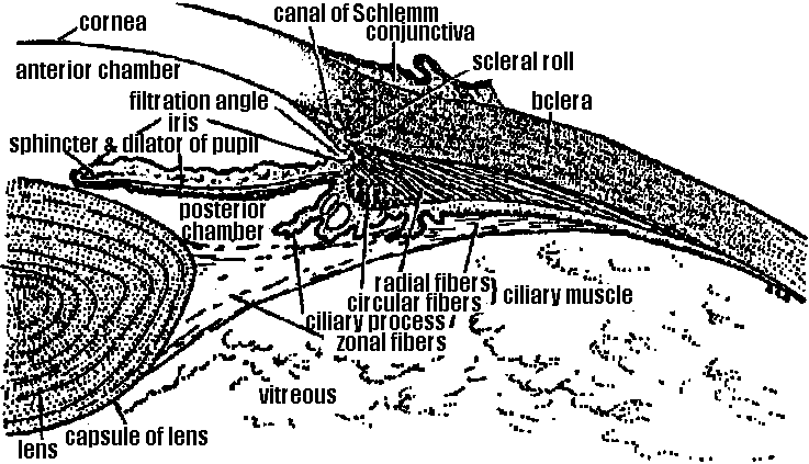 Fig. 7. Detail of anterior egnient of human eye. Ciliary pruccss has been distorted in cutting of section. Scleral roll is a narrow shelf of scieral tissue, on under side of which radial or izieridional fibers of ciliary muscle Originate. (Bloom & Fawcctt 1962). 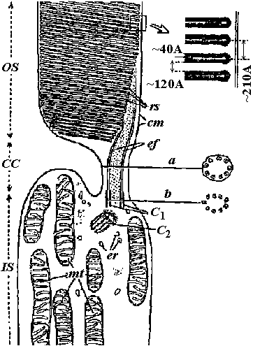 Fig. 17.4. Diagram to show the structure of a mammalian rod, as seen by electron microscopy. OS, outer segment; CC, connecting cilium: IS, inner segment; Cl and Cz, centrioles. Transverse sections through the connecting cilium (a) and the centriole (b) are shown at the right. rs, rod sacs; cf, ciliary filaments; cm, cell membrane; mi, mitochondrion; er, endoplasmic reticulum. (Robertis 1960)
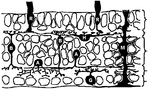 Outer nuclear layer Outer plexiform layer Inner nuclear layer Innter plexiform layer Ganglion cell layer Fig. 14.38. The principal types of cells and the layering of the vertebrate retina, based on mudpuppy (Necturus) retinas impregnated by the method of Golgi. R, receptor; H, horizontal cell; B. bipolar cells; A, amacrine cells; C, ganglion cell; and M, Muller (glial) cell. (Dowling 1970)
inner limiting membrane (内境界膜/板) Reproductive organ (生殖器)Dimorphism (二型性)Individual level: ♀ ↔ ♂Tissue level
ovary (卵巣) ↔ testis (精巣) |
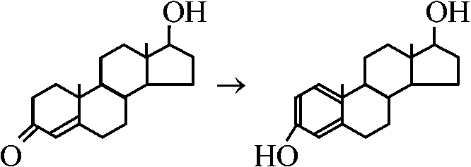
Gonads (生殖腺)Steroid hormones (ステロイドホルモン)acetate → 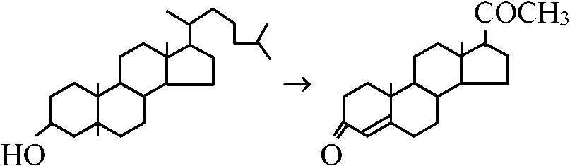 → →____________cholesterol____________progesterone
|
0. Muzzle (鼻口部)nose (鼻) =
external nose (nasus externus) (鼻軟骨) + 1. Nasal cavity (鼻腔)2. Paranasal sinuses (副鼻腔)3. Pharynx (咽頭)4. Larynx (喉頭)5. Trachea and bronchus (気管と気管支)6. Lung (肺)7. Pleura (胸膜) |
= digestive canal + appendages (glandula salivaria, liver, gall bladder, pancreas) Fig. Human digestive system |
Liver (肝臓)Tooth (歯, pl. teeth)Permanent, adult or second tooth (永久歯)Primary, baby or milk teeth (乳歯) Location and morphologyFront tooth or foretooth (前歯): 4 + 4
= Incisor (切歯) Back tooth (奥歯) = Posterior, grinding, cheek or wall teeth (臼歯) = premolar + molar
Premolar (小臼歯) 4 + 4 Third molar tooth(第三臼歯, 親知らず) 2 + 2 Total = 32 (adult)Dental formula (歯式)Classification of mammals based on the arrangement of teethEx. Human: two types of dental formula with aging
Primary teeth = I2/2 C1/1 M2/2 = 16 |
HistoryMüller, Johannes (1801-1858, German)First to observe cancer cells under a microscope 1838 Ueber den feineren Bau und die Formen der krankhaften Geschwülste
1840 On the nature and structural characteristics of cancer and of those morbid growths which may be confounded with it (translated version) 1858 proposed cellular theory of cancer (細胞学説)
cancer: caused by abnormal proliferation of cells, each derived from a pre‑existing cell, with tumor progression driven by cellular invasion and spread Cultured cell (培養細胞)Earle, Wilton R (dates unknown)1943 cultured fibroblasts (線維芽細胞) derived from mouse subcutaneous connective tissue
later called the L strain - milestone in mammalian cell culture |
Gey, George Otto (1899-1970) 1951 Hela cells (ヒーラ細胞)
the cervical cancer of Henrietta Lacks at Johns Hopkins Hospital Hela cells ⇒ isolated by Gey
established the first immortal human cell line for biomedical research Typical components include:
Amino acids (essential and non‑essential), inorganic salts, vitamins, carbohydrates (primarily glucose), a buffering system such as bicarbonate 1955 developed Eagle basal medium (BME)
rarely used today but remains important in the history of cell culture
improved from BME |